
A scleral contact lens (or “scleral lens,” “scleral contact,” ) is a large-diameter, rigid (gas-permeable) contact lens that vaults over the cornea and rests on the sclera (the white part of the eye).
Unlike corneal rigid gas-permeable (RGP) lenses or soft lenses that sit directly on the cornea, scleral lenses leave a fluid reservoir (typically sterile saline) between the posterior surface of the lens and the cornea, effectively “bridging” over the cornea.
This design provides a smooth optical interface, protection, and constant hydration to the corneal surface.
Over time, improvements in scleral lens materials, design software, diagnostic imaging, and fitting protocols have made them more accessible and successful.
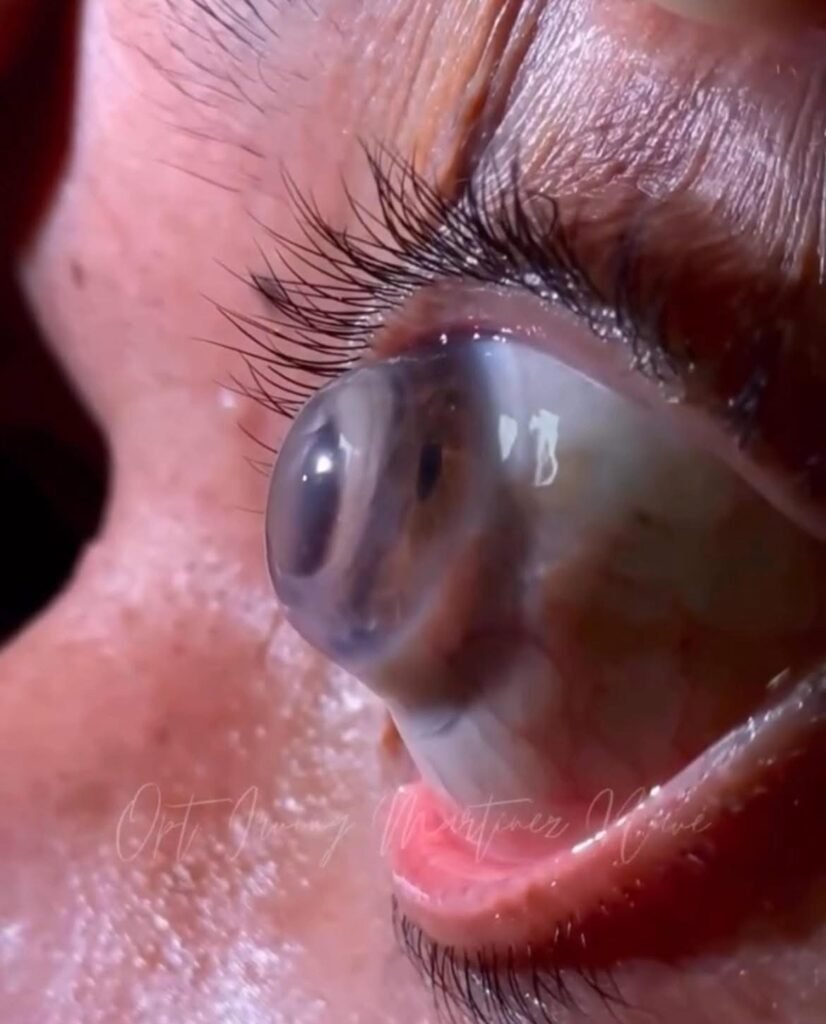
The Patient is wearing a Scleral contact lens. Image credit in Pic
Indications & Uses
Scleral lenses are used in various optical and therapeutic settings. Some of the main indications include:
- Irregular corneas / corneal ectasia
Conditions such as keratoconus, pellucid marginal degeneration, post-LASIK ectasia, or corneal scarring often result in irregular surfaces that are difficult to correct with spectacles or conventional lenses. Scleral lenses can mask irregular astigmatism and higher-order aberrations. - Ocular surface disease / dry eye
Because the fluid reservoir maintains hydration of the cornea, scleral lenses are used therapeutically for moderate to severe dry eye disease, neurotrophic keratitis, graft-versus-host disease (GVHD), Stevens-Johnson syndrome, and other ocular surface disorders. - Post-surgical / post-transplant cases
After corneal transplantation or other corneal surgery, residual irregular astigmatism or surface irregularities may remain. Scleral lenses may help in visual rehabilitation in such cases. - High refractive error / conventional lens failure
In some cases where spectacles or regular contact lenses fail (e.g. due to high astigmatism, intolerance, or lens decentration), scleral lenses are considered. - Therapeutic drug delivery / corneal healing support
Some advanced applications use scleral lenses as a vehicle to deliver drugs (e.g. cyclosporine) or support epithelial healing by maintaining a hydrated corneal environment (e.g. PROSE therapy).
How Scleral Lenses Help in Keratoconus
Scleral lenses are especially well suited for keratoconus because:
- Vaulting over the cornea: They create a tear-filled reservoir between the inner surface of the lens and the cornea, smoothing out the irregular corneal surface. This reduces issues like light scattering, ghosting, and distortion.
- Better visual acuity: Multiple studies show that patients with keratoconus obtain significantly better vision with well-fitted scleral lenses than with glasses or soft / conventional rigid lenses.
- Increased comfort: Because the lens rests on the less sensitive sclera (white of the eye) rather than the highly innervated, thin, and irregular cornea, patients usually find them more comfortable, especially when keratoconus is advanced or when there is corneal scarring
- Dryness relief / corneal protection: The fluid reservoir helps keep the cornea hydrated, protecting it from dryness and mechanical irritation from blinking or rubbing. This is very helpful because many keratoconus patients have coexisting ocular surface / tear film issues.
Outcomes in visual acuity are generally positive: multiple long-term studies show substantial gains in best-corrected visual acuity and improvement in patient-reported quality of life.
Basic Principles & Design Parameters
When designing or selecting a scleral lens, several geometric and material parameters must be considered:
- Diameter: Scleral lenses typically range from ~14.5 mm to over 24 mm. Lenses under ~18 mm are often called “mini-sclerals” or “semi-sclerals.”
- Sagittal depth / vault: The vertical height of the lens (or vault) is crucial to ensure clearance over the cornea and limbus without touching. This is often determined using corneal topography, optical coherence tomography (OCT), or scleral mapping.
- Landing or haptic zone: The peripheral portion of the lens that contacts the conjunctiva over the sclera must be designed to distribute pressure evenly and avoid impingement of blood vessels or conjunctival distortion. Variations include toric or asymmetric haptics, compression zones, or alignment designs.
- Edge lift / edge profile: This ensures exchange of tears beneath the lens and comfort at the edge. Too much lift can cause irritation; too little can impinge tissue or restrict tear flow.
- Oxygen permeability / material: Because the lens is thick and covers a wide area, the material’s oxygen transmissibility (Dk/t) is critical to corneal health. Modern materials attempt to optimize oxygen delivery.
- Fluorescein or dye use: Sometimes fluorescein or a yellow dye is mixed into the saline reservoir to help assess lens fit (vault, clearance, touch) under slit-lamp examination.
The Fitting Process: Step by Step
Fitting a scleral contact lens is more complex than fitting standard lenses — it is highly customized, iterative, and may require multiple visits. Below is a typical process:
- Preliminary evaluations & measurements
- Detailed ocular history and assessment of corneal health, surface disease, tear film, and eyelids.
- Topography / tomography of the cornea to detect irregularities, elevation, asymmetry.
- Measurement of scleral shape or scleral elevation profiles (if available). Some advanced clinics use scleral profilometry to map scleral shape.
- Determine target vault (a buffer margin over corneal apex and limbus).
- Selecting a trial lens / starting design
- Based on topography and fitting guides from lens manufacturers, the practitioner chooses an initial lens design (base curve, diameter, sagittal depth).
- Some fitters begin with an average K (keratometry) and choose a lens “flatter” than average to allow vaulting.
- Insertion & evaluation
- The practitioner fills the bowl of the lens with non-preserved sterile saline (often with fluorescein or dye).
- The lens is inserted while the patient fixates downward (practitioner sometimes assists with eyelids).
- After settling, evaluation using slit-lamp biomicroscopy, optical coherence tomography, or imaging to see the vault, clearance over the cornea & limbus, and peripheral fit.
- The practitioner checks for areas of contact (touch), inadequate vault, or conjunctival blanching or vascular compression.
- Adjustment & lens refinement
- Based on observations, modifications to sagittal depth, peripheral curves, or haptic alignment are made.
- The trial lens might be exchanged for a better-fitting design, and reevaluation is done. This may repeat several times until optimal fit is achieved.
- Comfort, insertion/removal ease, visual acuity, and lens stability are also evaluated.
- Final Rx & ordering custom lens
- Once the fit is acceptable, the practitioner finalizes the prescription and orders a custom lens from a laboratory.
- A “diagnostic protocol” may be used to guide the lab in fine adjustments.
- Follow-up & troubleshooting
- After patient receives the final lens, follow-up is essential to verify fit, comfort, vision, and ocular health.
- Common issues such as midday fogging (floaters or turbidity of fluid reservoir), decentration, discomfort, or conjunctival irritation are addressed via lens redesign. The SCOPE group has specifically investigated midday fogging phenomena.
- Periodic monitoring is required to ensure no adverse effects to the cornea (hypoxia, staining, vascularization).
Because this process is so customized, some practitioners may use tens of trial lenses; in simpler cases, only one or two may suffice.
Benefits & Advantages
Scleral lenses offer a number of advantages, especially in challenging cases:
- Improved vision & optical quality
By vaulting over the cornea, scleral lenses create a smooth refractive surface and reduce higher-order aberrations resulting from corneal irregularities.
Multiple studies demonstrate consistent improvement in best-corrected visual acuity. - Comfort & tolerance for sensitive eyes
Because they rest on less sensitive scleral tissue rather than the cornea, many patients unable to tolerate corneal lenses may do well with scleral lenses. - Continuous corneal hydration & therapeutic effect
The fluid reservoir baths the cornea during wear, offering relief for dry eyes and surface disease.
Scleral lenses are considered a therapeutic option in ocular surface disease management per DEWS II guidelines. - Protection & stability
The lens shields the cornea from environmental insult (dust, eyelid friction). The large diameter provides stability and resists dislodgement. - Versatility in difficult cases
They can often succeed where other lens modalities fail — e.g. post-surgical irregularities, corneal grafts, or severe dry eye. - Long-term performance
Studies show that the visual gains with scleral lenses are stable over years.
Challenges, Risks & Limitations
Scleral lenses are powerful tools, but fitting them and long-term wear carries risks and challenges:
- Hypoxia / corneal oxygenation
Because of lens thickness and coverage, oxygen delivery to the cornea may be limited. High Dk materials, thinner designs, and optimal tear exchange strategies help mitigate this. - Midday fogging / fluid reservoir turbidity
Debris or tear components may accumulate in the fluid reservoir, causing clouding or “fogging” of vision. This is one of the more common complaints and is an area of active research (e.g. SCOPE). - Insertion / removal difficulty
Because of their size, patients may struggle with handling the lens. Training, applicators, or suction plungers are often needed. - Conjunctival compression / vascular impingement
Poor haptic design may press on blood vessels, cause blanching, or lead to irritation. - Cost & access
Scleral lenses are more expensive than standard soft or corneal lenses, both for the lens itself and associated fitting visits. - Complex fitting required
Not all practitioners are experienced in scleral lens fitting; the time investment is greater. - Potential complications
These include corneal staining, epithelial trauma, neovascularization, or deposition. Close follow-up is essential.
Current Trends & Research
Some of the contemporary directions and findings in scleral lens research include:
- The SCOPE (Scleral Lenses in Current Ophthalmic Practice Evaluation) study tracks how scleral lenses are being used in practice, including lens designs, fitting habits, care regimens, and issues like midday fogging.Research is ongoing into quantifying midday fogging (optical density changes in fluid reservoir over time). Mayo Clinic
- Innovations in scleral shape mapping are helping to better customize haptics and reduce fit failures. reviewofcontactlenses.com
- Use of scleral lenses as drug delivery vehicles (e.g. in the PROSE platform) is being studied. ophthalmologytimes.com+1
- Bibliometric analysis shows the field of scleral lens research has expanded rapidly in recent years, with new hotspots in technology, materials, and fitting strategies. dovepress.com
Summary & Outlook
Scleral contact lenses represent a powerful and versatile modality for both vision correction and ocular surface therapy. Their ability to vault over the cornea and create a stable, hydrated environment makes them uniquely suited for challenging cases that are difficult or impossible to manage with conventional lenses or glasses.
However, success depends heavily on careful diagnostics, iterative fitting, and ongoing monitoring. The field continues to evolve, with advances in lens materials, diagnostic imaging, and fitting algorithms promising even better outcomes and broader accessibility.
If you like, I can also prepare a patient-friendly guide (simpler language) or a technical summary focusing on fitting protocols.
References & Suggested Reading
- “Update on indications, complications, and outcomes of scleral lenses.” PMC article. PMC
- “Current Trends in Scleral Lens Prescription, Management, and Evaluation.” PMC article. PMC
- “A Guide to Scleral Lens Fitting” (Boston materials) pi.bausch.com
- “Current applications and efficacy of scleral contact lenses.” PMC article. PMC
- “Therapeutic uses of scleral contact lenses for ocular surface disease.” PMC article. PMC
- “Mastering Scleral Lens Fitting: Lessons from 5,000 Patients.” Review of Contact Lenses article. reviewofcontactlenses.com
Fahmina is a qualified optometrist. She founded OptometrySkills.com to make professional-grade eye care knowledge accessible to practitioners and patients alike.