
Horseshoe Retinal Tear: Traction That Opens the Door to Retinal Detachment
A quiet flash of light, a sudden shower of floaters—these early warnings can easily be dismissed. Yet, behind them may lie a condition that demands immediate attention: the horseshoe retinal tear. This seemingly small retinal break carries a disproportionately high risk of progressing to retinal detachment if not treated promptly.
For clinicians, students, and patients alike, understanding this condition is essential
A horseshoe retinal tear (also called a flap tear) is a full-thickness break in the retina caused by vitreous traction.
What makes it distinctive is its U-shaped appearance, where a flap of retinal tissue remains attached at one end while the rest is pulled forward by the vitreous. This configuration is not just anatomical—it reflects an active, ongoing pulling force that keeps the tear open.
The development of a horseshoe tear is closely linked to age-related changes in the vitreous.
Posterior Vitreous Detachment (PVD)
As the eye ages, the vitreous gel liquefies and begins to separate from the retina—a process known as posterior vitreous detachment. While common and often harmless, problems arise when the vitreous remains strongly attached at certain نقاط (focal adhesion sites).
Persistent Vitreoretinal Traction
At these adhesion points, the vitreous does not detach smoothly. Instead, it pulls on the retina, creating mechanical stress. When this force exceeds the retinal strength, it results in a tear rather than a clean separation.
Formation of the Horseshoe Shape
The continued traction pulls the torn retinal tissue forward, forming the characteristic horseshoe (U-shaped) flap. Importantly, this flap remains mobile and attached at one edge—indicating that traction is still active.
Why Does a Horseshoe Retinal Tear Occur?
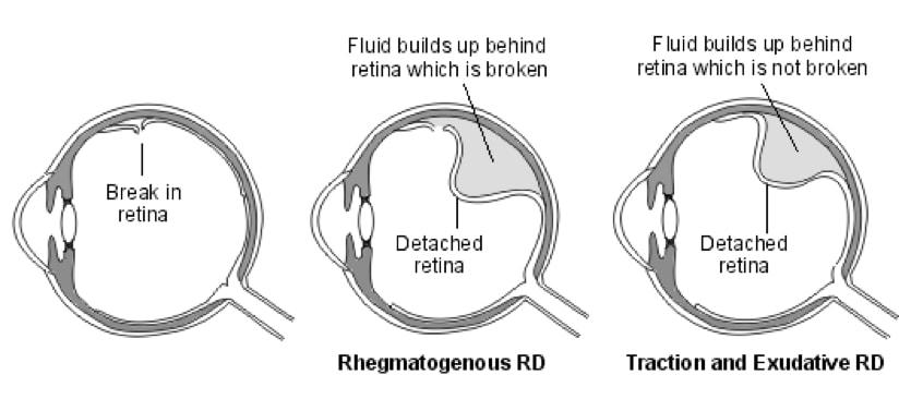
Once a tear forms, it creates a pathway for fluid movement.
- Liquefied vitreous enters through the tear
- Fluid accumulates in the subretinal space
- The neurosensory retina separates from the underlying retinal pigment epithelium
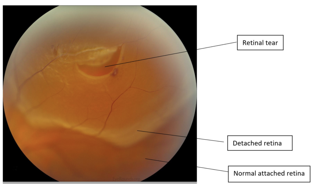
This process results in rhegmatogenous retinal detachment, the most common and vision-threatening form of retinal detachment.
Clinical Features of Horseshoe Retinal Tear
Symptoms Patients May Report
- Sudden flashes of light (photopsias)
- New onset or increase in floaters
- A shadow, curtain, or dark area in the visual field (late sign)
Signs on Examination
U-shaped retinal breakMobile flap attached at one endElevated edges due to tractionTypically located in the peripheral retina
Why Horseshoe Retinal Tears Are Clinically Significant
Not all retinal tears carry the same risk—but horseshoe tears are among the most dangerous.
- Persistent traction prevents spontaneous sealing
- The open flap allows continuous fluid entry
- Rapid progression to retinal detachment is possible
In clinical practice, these tears are considered high-risk lesions that often require urgent treatment.
Management of Horseshoe Retinal Tear
Laser Photocoagulation
The primary treatment involves applying laser burns around the tear:
- Creates a chorioretinal adhesion
- Forms a barrier to prevent fluid spread
- Reduces the risk of retinal detachment significantly
Cryotherapy
Used when laser is not feasible:
Observation and Follow-Up
- Selected asymptomatic cases may be monitored
- Requires strict follow-up to detect progression early
Key Concept to Remember
The danger of a horseshoe retinal tear lies not just in the break itself—but in the ongoing vitreoretinal traction that keeps it open and active.
Conclusion
A horseshoe retinal tear is a small lesion with potentially devastating consequences. It represents a dynamic process—where traction, fluid movement, and retinal separation interact rapidly.
The good news is that with early diagnosis and timely laser treatment, progression to retinal detachment can often be prevented entirely.
In ophthalmology, few interventions are as impactful as recognizing this tear early—because in this case, acting quickly preserves sight.
Fahmina is a qualified optometrist. She founded OptometrySkills.com to make professional-grade eye care knowledge accessible to practitioners and patients alike.