
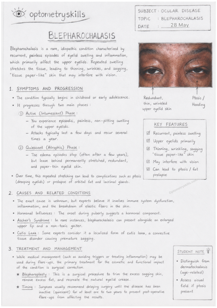
Blepharochalasis
Blepharochalasis is a rare eyelid disorder characterized by repeated episodes of painless eyelid swelling, mainly affecting the upper eyelids. Over time, these recurrent attacks stretch and weaken the delicate eyelid tissues, leading to thin, wrinkled, and sagging skin that resembles “tissue paper.” In severe cases, the condition may interfere with vision and cause cosmetic concerns.
This condition usually begins during childhood or early adolescence and progresses slowly through different stages. Although the exact cause remains unknown, inflammation and loss of elastic tissue are believed to play an important role.
What happens in Blepharochalasis?
Blepharochalasis is an uncommon condition involving recurrent edema (swelling) of the eyelids. Each episode of swelling damages the elastic fibers within the eyelid skin. As a result, the eyelids gradually become loose, atrophic, and redundant.
The upper eyelids are most commonly affected, though bilateral involvement may occur. Repeated inflammation may eventually lead to complications such as ptosis, prolapse of orbital fat, or lacrimal gland displacement.
Symptoms of Blepharochalasis
Patients with blepharochalasis may present with the following symptoms:
- Recurrent painless swelling of the eyelids
- Thin, wrinkled, “tissue paper-like” skin
- Sagging upper eyelids
- Ptosis (drooping eyelids)
- Eyelid discoloration or visible blood vessels
- Heaviness around the eyes
- Visual obstruction in severe cases
The swelling episodes are usually non-pitting and may last for a few days before resolving spontaneously.
Stages of Blepharochalasis
1. Active (Intumescent) Phase
This phase is characterized by recurrent attacks of eyelid swelling. The episodes occur intermittently and may recur several times a year. During this stage:
- Swelling is painless
- The upper eyelids are primarily involved
- Inflammation stretches the eyelid tissue
- Episodes may last from hours to days
2. Quiescent (Atrophic) Phase
After repeated attacks, the disease enters a more stable stage where active swelling decreases or stops completely. However, permanent tissue damage remains.
Features of this phase include:
- Thin and atrophic eyelid skin
- Wrinkling and redundancy
- Ptosis
- Orbital fat prolapse
- Lacrimal gland prolapse
Causes of Blepharochalasis
The exact cause of blepharochalasis is still unknown, making it an idiopathic condition. However, several mechanisms have been suggested:
Inflammatory Changes
Repeated inflammatory episodes damage the elastic fibers in the eyelid skin, causing progressive tissue laxity.
Immune System Dysfunction
Some researchers believe immune-mediated inflammation contributes to recurrent swelling.
Hormonal Influence
The onset during puberty suggests hormonal factors may play a role in disease development.
Associated Conditions
Ascher’s Syndrome
Blepharochalasis may occur as part of Ascher’s syndrome, which includes:
- Blepharochalasis
- Double upper lip
- Non-toxic thyroid enlargement (goiter)
Cutis Laxa
Some experts consider blepharochalasis a localized form of cutis laxa, a connective tissue disorder associated with loose, sagging skin.
Diagnosis
Diagnosis is mainly clinical and based on patient history and examination findings. Important features include:
- Recurrent eyelid edema
- Atrophic and wrinkled eyelid skin
- Ptosis
- Eyelid laxity
The condition must be differentiated from dermatochalasis, allergic eyelid edema, angioedema, and inflammatory eyelid diseases.
Treatment of Blepharochalasis
Medical Management
There is no definitive medical cure for blepharochalasis. During active episodes, management may include:
- Avoiding triggers
- Managing inflammation
- Observation during mild attacks
Medical treatment is usually limited because the condition is primarily structural after repeated episodes.
Surgical Management
Blepharoplasty
Blepharoplasty is the main treatment option for patients with functional or cosmetic concerns.
The procedure may involve:
- Removal of excess eyelid skin
- Correction of ptosis
- Removal of prolapsed orbital fat
- Reconstruction of the eyelid crease
Timing of Surgery
Surgery is generally delayed until the disease has remained inactive for at least 1–2 years. Performing surgery during the active stage may lead to recurrence and poor surgical outcomes.
Prognosis
The condition often stabilizes after several years, but the tissue changes may remain permanent. Surgical correction usually provides good cosmetic and functional improvement once the disease becomes quiescent.
Conclusion
Blepharochalasis is a rare but important eyelid disorder that causes recurrent swelling and progressive thinning of the eyelid skin. Repeated inflammatory episodes eventually result in sagging, wrinkling, and ptosis. Early recognition and proper differentiation from other eyelid conditions are essential. While medical therapy has limited benefit, surgical correction during the inactive stage can significantly improve both appearance and visual function.

Optometry education for Optometry students and eye care professionals. Managed by qualified optometrists. For informational purposes only — not a substitute for professional eye care.