
Amsler grid eye test Printable and Online: Amsler Grid: A Simple Yet Powerful Tool to Detect Macular Problems Early
In clinical practice, some of the most serious vision-threatening conditions begin quietly—without pain, without obvious warning. The Amsler Grid is one of those beautifully simple tools that allows both clinicians and patients to see what the eye is silently experiencing. A small grid, a central dot, and a few seconds of attention can reveal distortions that may otherwise go unnoticed until significant damage has occurred.
What is the Amsler Grid?


The Amsler Grid is a central visual field test designed to assess the central 20° of vision, particularly the macular region.
- It consists of a 10 cm square grid
- Each small square subtends:
- 1° in standard charts
- 0.5° in fine grids (Chart 7 – more sensitive)
- A central fixation dot helps maintain steady gaze
This test is widely used for:
- Screening macular disease
- Monitoring progression (e.g., AMD, CNV)
- Detecting subtle central field defects
Clinical Significance
The Amsler Grid is primarily used to detect:
- Metamorphopsia (distorted/wavy lines) → often seen in macular disease
- Scotomas (missing or blank areas) → may indicate optic nerve or retinal pathology
- Central visual field defects
Clinical Insight:
- Macular disease: lines appear wavy or distorted
- Optic neuropathy: lines appear faint or missing, not distorted
Step-by-Step Technique (Clinical Standard)
To ensure accuracy, the technique must be followed carefully:
Preparation
- Pupils should not be dilated
- Avoid prior slit-lamp examination (prevents photostress effect)
- Ensure:
- Good illumination
- Proper refractive correction (especially for presbyopia)
- Viewing distance ~ 33 cm
Procedure
- Cover one eye
- Ask the patient to fixate on the central dot
- While maintaining fixation, ask:
- Do any lines appear wavy or distorted?
- Then ask:
- Are there any blurred or missing areas?
- Confirm:
- Can the patient see all four corners and borders?
Important Interpretation Clue
- Missing corners or borders may suggest:
- Glaucoma
- Retinitis pigmentosa
- Not just macular pathology
Recording
Patients can:
- Mark abnormalities on a printed grid
- Track changes over time
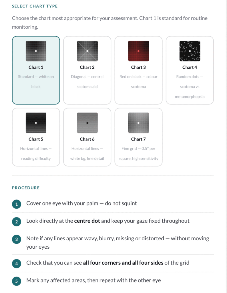
Types of Amsler Grid Charts


There are seven variations, each serving a specific diagnostic purpose:
Chart 1 – Standard Grid
- White grid on black background
- 400 small squares (5 mm each)
- Basic screening tool
Chart 2 – Fixation Aid
- Includes diagonal lines
- Helps patients with central scotoma maintain fixation
Chart 3 – Red Grid
- Stimulates long-wavelength (red) cones
- Detects:
- Colour scotomas
- Toxic maculopathy
- Optic nerve/chiasmal issues
Chart 4 – Random Dots
- No structured lines
- Helps differentiate:
- Scotoma vs metamorphopsia
Chart 5 – Horizontal Lines
- Focus on specific meridians
- Useful in:
- Patients with reading difficulty
Chart 6 – Enhanced Horizontal Grid
- White background
- Closer central lines
- Provides higher sensitivity
Chart 7 – Fine Central Grid
- Smaller squares (0.5°)
- More sensitive for subtle defects
Home Monitoring: A Powerful Preventive Tool
Patients at risk of choroidal neovascularization (CNV) or macular disease should be encouraged to use the Amsler Grid at home.
Why this matters:
- Early detection = better prognosis
- Changes can be reported before vision loss progresses
Amsler eye test grid Online tool
Fahmina is a qualified optometrist. She founded OptometrySkills.com to make professional-grade eye care knowledge accessible to practitioners and patients alike.