
Last updated:
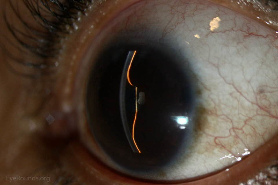
Iris bombe is a forward ballooning of the iris caused by absolute pupillary block. It is a classic secondary cause of angle-closure glaucoma and a common examination topic because the mechanism, signs, and treatment follow a clear sequence.
Definition
Iris bombe (also written iris bombé) is anterior bowing of the iris between the pupil margin and the iris root. It occurs when the pupil is sealed, usually by 360° posterior synechiae (seclusio pupillae), so aqueous cannot pass from the posterior chamber into the anterior chamber.
The name comes from the French bombé, meaning convex or bulging. The iris is pushed forward like a dome by fluid trapped behind it.
Normal aqueous flow
- Aqueous is produced by the ciliary body and enters the posterior chamber.
- It passes through the pupil into the anterior chamber.
- It leaves the eye mainly through the trabecular meshwork and Schlemm canal.
Anything that completely blocks step 2 can produce iris bombe.
Pathophysiology
- Inflammation, trauma, or surgery causes the pupil margin to stick to the anterior lens capsule (posterior synechiae).
- If adhesions become circumferential, the pupil is secluded. Aqueous cannot move forward.
- Pressure rises in the posterior chamber relative to the anterior chamber.
- The iris, fixed at the pupil margin and at the root, bows forward in between.
- The peripheral iris is pushed against the cornea and trabecular meshwork.
- The angle closes, first by apposition and later by peripheral anterior synechiae (PAS).
- Intraocular pressure (IOP) rises. This is secondary angle-closure glaucoma.
This is absolute pupillary block, not the relative pupillary block of primary angle closure. In primary angle closure the pupil is not sealed by synechiae; resistance is only from iris–lens contact in a predisposed eye.
Uveitic eyes often have more than one pressure mechanism at once. Iris bombe may coexist with trabecular inflammation, PAS, steroid-related pressure rise, or damage to the outflow pathway. Flattening the iris does not always normalize IOP.
Causes
The usual setting is anterior uveitis severe or prolonged enough to form posterior synechiae. Other recognized settings include:
- Chronic or recurrent iridocyclitis
- Trauma
- Intraocular surgery
- Prolonged postoperative inflammation
- A pupil occluded by a fibrous or fibrovascular membrane (occlusio pupillae), which may accompany seclusio pupillae
Sectoral bowing can occur with broad but incomplete synechiae. The full, circumferential dome is the classic picture of complete seclusio pupillae.
Clinical features
patients may already be known to have uveitis. Presentation can be subacute or acute.
- Ocular pain and headache
- Redness, often ciliary injection
- Blurred vision
- Halos if the cornea is edematous
- Nausea and vomiting if IOP is very high
- A history of a sticky, poorly dilating, or irregular pupil
Examination
| Finding | What it means |
|---|---|
| Irregular or fixed pupil | Posterior synechiae |
| 360° pupil–lens adhesion | Seclusio pupillae |
| Forward convex iris | Iris bombe |
| Very shallow peripheral anterior chamber | Iris apposed to cornea |
| Relatively preserved central chamber | Lens has not moved forward as a whole |
| High IOP | Secondary angle closure |
| corneal epithelial edema | Acute pressure rise |
| Keratic precipitates, cells, flare | Underlying or recent uveitis |
| Closed angle on gonioscopy | Appositional closure and/or PAS |
Useful bedside points:
- Van Herick estimation shows a very narrow or absent peripheral chamber.
- The iris has a smooth, domed contour rather than a flat plane.
- Attempted dilation shows where the iris remains stuck to the lens.
- Gonioscopy is essential. Indentation gonioscopy helps separate appositional closure, which may open, from PAS, which will not.
- Corneal edema may hide the angle until pressure is lowered.
Anterior segment OCT or ultrasound biomicroscopy, when available, shows the bowed iris, a sealed pupil, and a closed peripheral angle. These tests support the clinical diagnosis; they are not required to recognize the classic picture.
Differential diagnosis
The important comparison is with other causes of a shallow chamber and high pressure.
| Condition | Chamber | Key distinction |
|---|---|---|
| Iris bombe | Periphery shallow; center relatively deeper; pupil stuck to lens | History of uveitis or synechiae; domed iris |
| Primary angle closure | Periphery shallow; center usually formed | No seclusio pupillae; narrow-angle anatomy; relative pupillary block |
| Aqueous misdirection (malignant glaucoma) | Central and peripheral chambers both very shallow | Whole lens–iris diaphragm pushed forward, often after surgery |
| Choroidal effusion or hemorrhage | Shallow chamber, often with pain | Posterior segment or B-scan shows choroidal elevation |
| Plateau iris | Periphery narrow after iridotomy | Iris root configured anteriorly; pupil not sealed |
In iris bombe the lens is in a normal position and the pupil margin is tethered to it. In aqueous misdirection the lens and iris move forward together, so the central chamber is shallow as well.
Complications
- Acute or chronic glaucomatous optic neuropathy
- Permanent PAS and chronic angle-closure glaucoma
- Corneal edema and, if prolonged, corneal endothelial damage
- Cataract
- Recurrent closure if a laser opening seals with fibrin
- Mixed-mechanism uveitic glaucoma even after the iris flattens
Management principles
Treatment has three aims: lower IOP, equalize pressure across the iris, and control inflammation. Doses are not listed here because they depend on the patient and local protocols.
Lower the pressure
- Topical aqueous suppressants: beta-blockers, alpha-2 agonists, and carbonic anhydrase inhibitors, if not contraindicated
- Systemic carbonic anhydrase inhibitors for a large acute rise
- Hyperosmotic agents in selected acute, vision-threatening rises
- Avoid routine use of miotics. The pupil is already sealed, and miotics do not relieve this block; they may worsen inflammation and synechiae
- Prostaglandin analogues are generally avoided while inflammation is active
Medical treatment protects the optic nerve and clears the cornea. It does not remove the pupillary block.
Bypass the block
Definitive treatment is an opening in the iris so aqueous can pass directly from the posterior to the anterior chamber.
- Laser peripheral iridotomy, usually Nd:YAG, when the cornea is clear enough
- Surgical iridectomy if the view is poor, laser fails, or the opening is likely to close
- In active uveitis the opening may seal with fibrin, so a sufficiently large opening, close review, and sometimes a second procedure are needed
- After a successful opening the iris should flatten if closure was mainly appositional
If extensive PAS are already present, iridotomy or iridectomy may flatten the iris but not control IOP. Filtering surgery, a drainage device, or other glaucoma surgery may then be required, timed with control of inflammation. Cataract surgery is sometimes part of later management, but it is not the emergency treatment of the block itself.
Treat the inflammation
- Intensive topical corticosteroid for active anterior uveitis, unless a specific contraindication exists
- Cycloplegia is used in anterior uveitis to relieve pain and reduce formation of new posterior synechiae
- Continue uveitis treatment after the iris opening, because ongoing inflammation can close the iridotomy and damage trabecular outflow
Follow-up
- Recheck IOP, anterior chamber depth, iris contour, patency of the opening, and inflammation
- Repeat gonioscopy to see how much of the angle has reopened
- Assess the optic disc and visual field once the acute episode allows
- Investigate and treat the underlying uveitis when a systemic association is suspected
Fellow-eye laser iridotomy is not automatic. That prophylaxis belongs to primary angle-closure disease. The other eye is managed according to its own inflammation, synechiae, and angle anatomy.
Prevention
In anterior uveitis, early cycloplegia and mydriasis are used to keep the pupil moving and reduce posterior synechiae. Breaking fresh synechiae before they become circumferential is the practical way to prevent iris bombe. Patients with recurrent uveitis need prompt review if pain, redness, or visual change returns.
High-yield revision points
- Iris bombe = seclusio pupillae → absolute pupillary block → forward iris dome → secondary angle closure.
- Classically follows anterior uveitis, not a narrow angle alone.
- Peripheral chamber is shallow; central chamber is relatively spared. Uniform central shallowing suggests aqueous misdirection or a posterior pushing force.
- Gonioscopy distinguishes appositional closure from PAS.
- Miotics do not treat the block.
- Pressure control is temporary; an iridotomy or iridectomy bypasses the block.
- Inflammation must be treated or the opening and the angle may fail.
- IOP may stay high after the iris flattens because uveitic glaucoma is often multifactorial.
- Do not apply primary angle-closure fellow-eye rules automatically.
One-line summary
Iris bombe is inflammatory or post-inflammatory sealing of the pupil that traps aqueous behind the iris, bows the iris forward, closes the angle, and needs urgent pressure control, an iris opening, and treatment of the underlying uveitis.

Optometry education for Optometry students and eye care professionals. Managed by qualified optometrists. For informational purposes only — not a substitute for professional eye care.