
Last updated:
Haab’s Striae Notes
Index
Definition & Clinical Significance
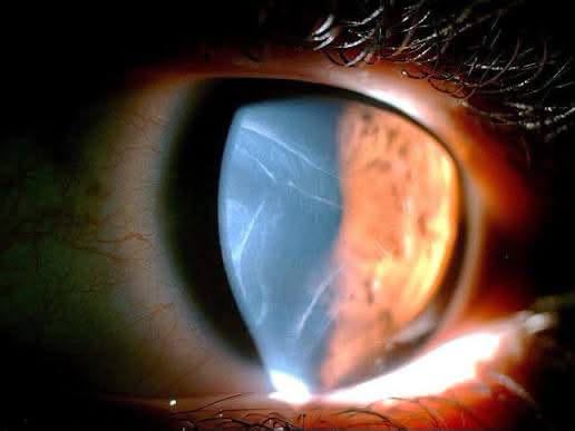
- Haab’s striae are horizontal, curvilinear, or concentric breaks in Descemet’s membrane (DM) caused by mechanical stretching of the infant cornea under high intraocular pressure.
- They are a hallmark sign of primary congenital glaucoma (PCG) and other forms of infantile-onset glaucoma(e.g., secondary congenital glaucoma, glaucoma associated with congenital anomalies or anterior segment dysgenesis).
- Their presence indicates significantly elevated intraocular pressure (IOP) during the first 3 years of life, when the sclera and cornea are still elastic, pliable, and prone to stretching.
Pathophysiology
- Elevated IOP in infants causes corneal enlargement (buphthalmos) due to the inherent elasticity of the young cornea.
- The Descemet’s membrane is a non-elastic basement membrane secreted by corneal endothelial cells.
- As the cornea stretches, DM ruptures along lines of stress, creating Haab’s striae.
- Why horizontal?
- The cornea stretches more in the horizontal meridian due to the elliptical shape of the infantile cornea and the distribution of stress forces.
- The endothelium migrates over the breaks and lays down new DM, creating a double-contoured appearance on histology.
Clinical Features
| Feature | Description |
|---|---|
| Appearance | Horizontal or curvilinear, glass-like lines on the posterior cornea. |
| Location | Typically central or paracentral, sparing the periphery. |
| Number | Usually multiple, but a single striae may be seen in mild cases. |
| Associated Signs | – Buphthalmos (enlarged cornea, >12 mm diameter in newborns). – Corneal edema (due to endothelial dysfunction). – Photophobia, blepharospasm, epiphora (classic triad of congenital glaucoma). |
| Age of Onset | <3 years (cornea loses elasticity after this age). |
Differential Diagnosis of Descemet’s Membrane Breaks & Enlarged Cornea
| Condition | Key Distinguishing Features |
| Birth Trauma (Forceps Injury) | – Vertical or oblique DM breaks (due to compression of the globe against the orbital rim). – Unilateral (frequently left eye due to Left Occiput Anterior birth presentation). – Normal corneal diameter (no buphthalmos) and normal IOP. |
| Megalocornea | – Bilateral enlarged cornea ($>13\text{ mm}$) without IOP elevation or Haab’s striae. – X-linked recessive; clear cornea, normal cup-to-disc ratio, normal anterior segment. |
| Corneal Dystrophies (e.g., CHED) | – Congenital Hereditary Endothelial Dystrophy: Diffuse, bilateral corneal edema from birth. – No DM breaks, normal corneal diameter, normal IOP. |
| High Axial Myopia | – Increased axial length without corneal stretching or DM breaks. – Normal IOP and normal corneal diameter. |
Diagnostic Workup
- History & Examination
- Assess for photophobia, tearing, blepharospasm.
- Measure corneal diameter (normal newborn: 9.5–10.5 mm; >12 mm suggests buphthalmos).
- Slit-lamp exam: Haab’s striae appear as refractile lines on the posterior cornea.
- IOP Measurement
- Use Tonopen or Perkins tonometer under anesthesia (avoid general anesthesia if possible in infants).
- Normal IOP in infants: ~10–15 mmHg; >20 mmHg is suspicious.
- Gonioscopy
- Evaluate the anterior chamber angle for trabeculodysgenesis (e.g., flat iris insertion, Barkan’s membrane).
- Axial Length & Pachymetry
- Increased axial length and thin central cornea (due to stretching).
- Imaging
- UBM (ultrasound biomicroscopy) or AS-OCT to assess angle structures.
Management
1. Medical Therapy (Temporizing Bridge to Surgery)
Primary congenital glaucoma is essentially a surgical disease. Medical therapy is used only to temporarily lower IOP prior to surgery or as adjunctive therapy in refractory cases.
- Topical beta-blockers (e.g., timolol 0.25%) or carbonic anhydrase inhibitors (e.g., dorzolamide).
- Avoid: Prostaglandin analogs (ineffective in infants) and miotics (paradoxical IOP rise).
🛑 CRITICAL SAFETY CONTRAINDICATION:
Alpha-2 Agonists (e.g., Brimonidine, Apraclonidine) are STRICTLY CONTRAINDICATED in infants and young children
2. Surgical Therapy (Definitive Treatment)
| Procedure | Indication & Approach |
| Goniotomy | – First-line option when the cornea is clear enough to visualize angle structures. – Ab-interno incision of the trabecular meshwork under direct gonioscopic visualization. |
| Trabeculotomy (Incisional or 360° GATT) | – First-line option when corneal opacity/edema obscures the anterior chamber angle. – Ab-externo cut into Schlemm’s canal to cleave the trabecular meshwork into the anterior chamber. |
| Combined Trabeculotomy + Trabeculectomy (CTT) | – Preferred in severe presentations, advanced buphthalmos, or developing regions where single interventions carry lower success rates. |
| Glaucoma Drainage Devices / Cyclophotocoagulation | – Reserved for refractory cases after failed primary angle surgeries (e.g., Ahmed or Baerveldt implants). |
Postoperative Care & Long-Term Rehabilitation
- Regular Monitoring: Lifelong follow-up of IOP, corneal diameter, axial length, optic nerve cupping, and visual acuity.
- Refractive Error Correction: Haab’s striae and axial elongation cause high astigmatism and axial myopia. Prompt optical correction (glasses/contact lenses) is required.
- Amblyopia Management: Occlusion therapy (patching) for anisometropic or deprivation amblyopia.
Prognosis & Complication
Prognosis
- Early diagnosis & surgery: 80–90% success in IOP control.
- Poor prognostic factors:
- Late presentation (>1 year).
- Severe corneal edema or buphthalmos.
- Associated ocular anomalies (e.g., aniridia, Sturge-Weber syndrome).
- Visual outcomes depend on:
- Duration of elevated IOP before treatment.
- Optic nerve damage at presentation.
- Amblyopia (due to corneal opacities or anisometropia)
Long-Term Complications
Complications
- Corneal
- Persistent edema (if endothelial damage is severe).
- Band keratopathy (chronic edema).
- Optic Nerve
- Glaucomatous cupping (irreversible if untreated).
- Refractive
- High myopia (due to axial elongation).
- Anisometropia (if unilateral).
- Amblyopia
- Deprivation amblyopia (corneal opacity) or anisometropic amblyopia.
Key High-Yield Exam Points
- ✅ Haab’s Striae = Breaks in Descemet’s Membrane due to horizontal stretching in infantile glaucoma.
- ✅ Horizontal/Concentric Orientation (vs. Vertical/Oblique in forceps birth trauma).
- ✅ Classic Symptom Triad of PCG: Epiphora, Photophobia, Blepharospasm.
- ✅ Surgical Choice: Goniotomy (Clear cornea) vs. Trabeculotomy (Cloudy cornea).
- ✅ Black Box Contraindication: Brimonidine is contraindicated in infants (causes CNS depression and apnea).
- ✅ Primary Cause of Permanent vision Loss: Amblyopia (anisometropic or deprivation).
High-Yield Mnemonics
- “Haab’s = Horizontal” (vs. Vertical in Forceps Birth Trauma).
- “3 P’s of PCG Symptoms”: Photophobia, Palpebral spasm (blepharospasm), Profuse tearing (epiphora).
- “3 H’s of Physical Examination”: Haab’s striae, Hazy cornea, Huge eye (buphthalmos).
Primary References
- American Academy of Ophthalmology (AAO): Basic and Clinical Science Course (BCSC), Section 6: Pediatric Ophthalmology and Strabismus.
- American Academy of Ophthalmology (AAO): Basic and Clinical Science Course (BCSC), Section 10: Glaucoma.
- Beck, A. D., et al. Childhood Glaucoma. Childhood Glaucoma Research Network (CGRN) Classification.
- Becker-Shaffer’s Diagnosis and Therapy of the Glaucomas (8th Edition).

Optometry education for Optometry students and eye care professionals. Managed by qualified optometrists. For informational purposes only — not a substitute for professional eye care.