
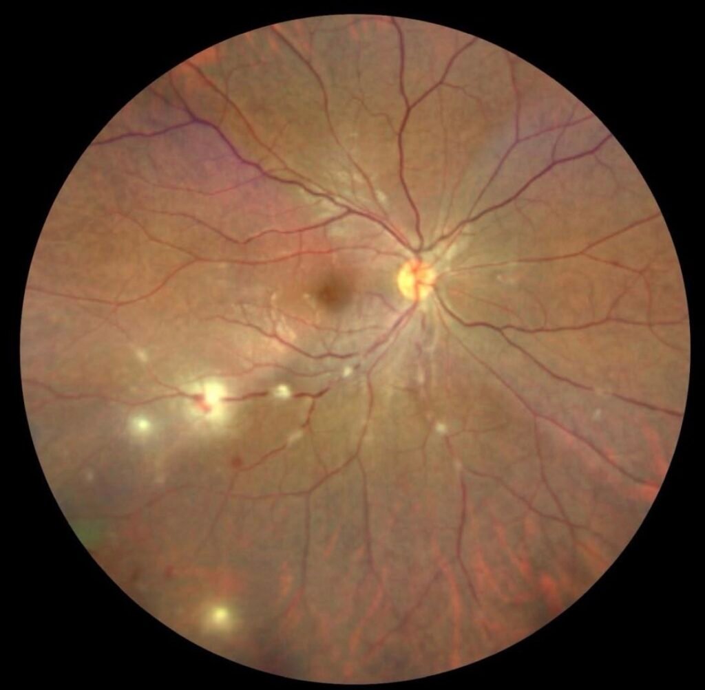
Understanding Tubercular Retinitis and Retinal Vasculitis: An Ocular TB Case
Tuberculosis is commonly associated with lung disease, but what many people do not realize is that TB can also affect the eyes in complex and sight-threatening ways. One of the most challenging presentations is tubercular retinitis with retinal vasculitis, where inflammation affects the retina and retinal blood vessels.
Recently, I studied a very interesting retinal case shared by the European VitreoRetinal Society (EVRS), and it highlights how important careful diagnosis and follow-up are in ocular inflammatory disease.
Case Presentation
A 22-year-old male presented with a one-week history of blurred vision in the left eye. Despite the symptoms, visual acuity remained relatively good at 20/20 in the right eye and 20/25 in the left eye.
However, retinal examination revealed something much more significant.
Color fundus photography showed multifocal areas of retinitis along with retinal phlebitis in both eyes. These findings raised suspicion for an inflammatory or infectious retinal vasculitis.
An important clue came from the patient’s family history. His sister had previously undergone treatment for pulmonary tuberculosis. She also had Takayasu arteritis requiring renal artery embolization and was on multiple immunosuppressive medications.
Investigations and Diagnosis
Further systemic investigations were performed.
Chest CT imaging revealed paratracheal and subcarinal lymph node calcification, suggesting previous or latent tuberculosis exposure. Mantoux skin testing was also strongly positive.
Based on the retinal findings, systemic history, imaging, and supportive laboratory evidence, the patient was diagnosed with presumed ocular tuberculosis presenting as tubercular retinitis and retinal vasculitis.
Treatment
The patient was started on anti-tubercular therapy (ATT) along with oral corticosteroids to control inflammation.
Initially, the response was encouraging.
After three weeks, visual acuity improved to 20/20 in both eyes, and the retinal inflammation had reduced significantly.
However, an important development occurred during follow-up.
Despite overall improvement, a new area of active inflammation appeared inferonasally in the right eye. This demonstrates how ocular TB can remain active even while treatment appears to be working clinically.
Two weeks later, the retinitis and vasculitis became inactive in both eyes, although residual retinal hemorrhages were still visible in the left eye. Steroid tapering was then started carefully.
Why This Case Is Important
This case teaches several important clinical lessons.
1. Ocular TB Can Mimic Many Diseases
Tuberculosis involving the eye can resemble multiple inflammatory conditions including:
- Anterior uveitis
- Intermediate uveitis
- Posterior uveitis
- Panuveitis
- Retinal vasculitis
- Neuroretinitis
- Choroiditis
- Optic neuropathy
Because of this, diagnosis can be difficult and requires careful correlation between ocular findings and systemic investigations.
2. Diagnosis Is Often Presumed, Not Definitive
In many ocular TB cases, direct microbiological confirmation is difficult. Therefore, diagnosis is usually based on:
- Clinical examination
- Imaging findings
- Tuberculosis exposure history
- Mantoux or IGRA testing
- Response to therapy
Laboratory tests support the diagnosis but should not be the sole deciding factor.
3. Close Follow-Up Is Essential
One of the most important aspects of this case is that a new inflammatory lesion developed even after initial improvement.
This emphasizes that retinal vasculitis patients require close and continuous monitoring throughout treatment.
Final Thoughts
Tubercular retinal vasculitis remains highly relevant in endemic countries such as India and other regions where tuberculosis exposure is common.
Cases like this remind us how interconnected systemic disease and ocular findings can be. They also highlight the importance of detailed retinal examination, multimodal investigation, and careful long-term follow-up.
As an optometry student and eye care learner, studying cases like this is incredibly valuable for understanding real-world retinal disease presentation and management.
Image Credits:
@retina.rocks

Optometry education for Optometry students and eye care professionals. Managed by qualified optometrists. For informational purposes only — not a substitute for professional eye care.