
Soemmering’s Ring: Formation, Clinical Significance, and Optometric Considerations
Soemmering’s ring is a well-recognized postoperative anatomical change that develops in the capsule after cataract extraction. Although usually asymptomatic, it holds clinical significance for optometrists, especially when evaluating pseudophakic patients presenting with visual disturbances, posterior capsular opacification (PCO), or IOL-related issues. Understanding its formation and appearance helps clinicians differentiate normal postoperative findings from pathology.
What Is Soemmering’s Ring?
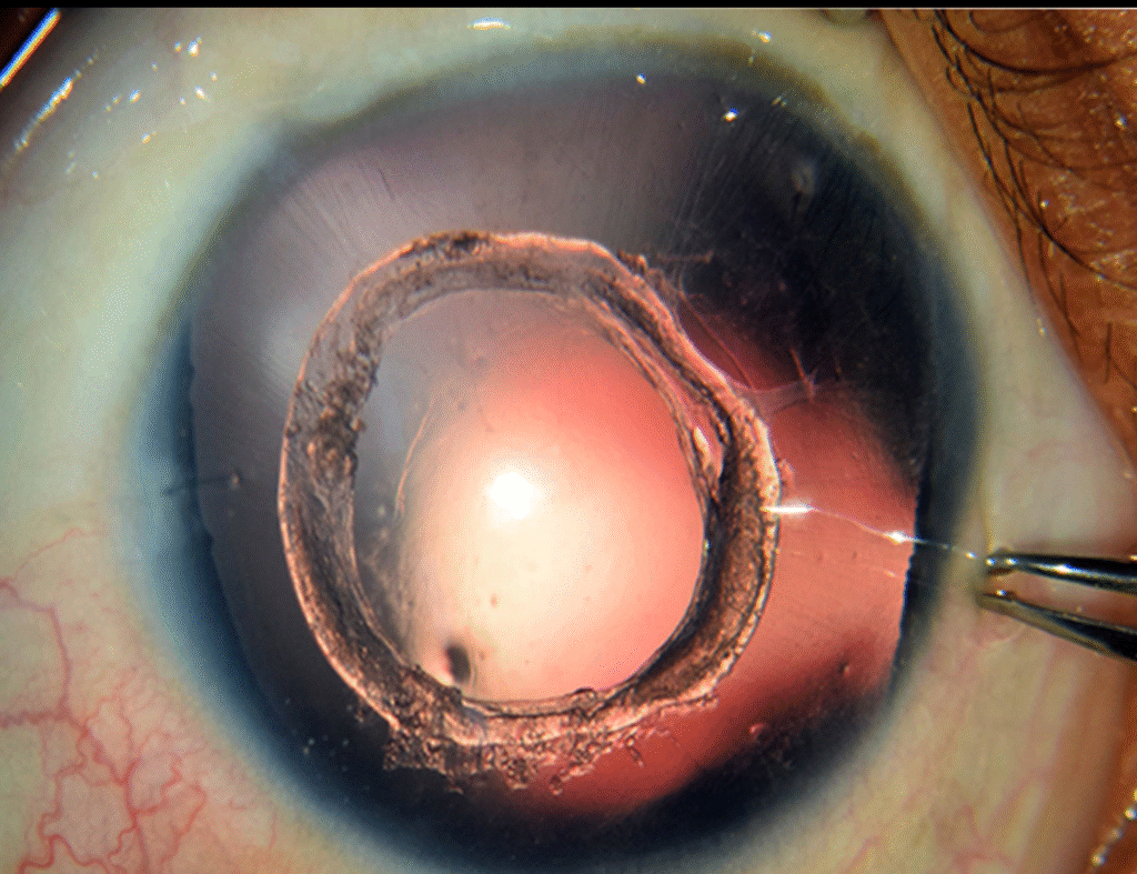
Soemmering’s ring is an annular (ring-shaped) accumulation of residual equatorial lens epithelial cells (LECs) and cortical fibers that remain after cataract surgery.
These cells proliferate and migrate along the equatorial region of the lens capsule, forming a dense, whitish, donut-shaped ring between:
- the anterior capsule remnant
- the posterior capsule
This structure is typically located behind the iris plane, making it visible on slit-lamp examination in a dilated eye.
Image credit- aao journal
How Does Soemmering’s Ring Form?
The formation is driven by two key mechanisms:
1. Residual Lens Epithelial Cell Proliferation
Even with modern surgical techniques, some LECs remain in the equatorial region of the capsule. These cells continue to metabolize and divide postoperatively.
2. Cortical Fiber Regeneration
The proliferating LECs differentiate into new cortical lens fibers. These fibers accumulate circumferentially around the equator, forming a ring.
Why a Ring Shape?
The centre of the capsule is cleaned during surgery, but the equator is harder to access, so any remaining cells proliferate around that region, creating a symmetrical ring.
Clinical Appearance
On slit-lamp, Soemmering’s ring appears as:
- A white or yellowish circular mass
- Behind the iris and outside the visual axis
- Between anterior and posterior capsule remnants
- Best observed with dilation and retroillumination
Potential Complications
1. Capsular Bag Distension
The ring can enlarge and push the capsule forward, potentially altering IOL position.
2. IOL Decentration or Tilt
Large rings may mechanically distort the capsular bag, affecting IOL stability.
3. Pigment Dispersion / Iris Chafing
In some patients, the ring may mechanically contact the iris.
4. Glaucoma Risk (rare)
If capsular distention leads to angle crowding.
5. Complication During Secondary IOL Surgery
The presence of a ring may complicate sulcus placement or bag manipulation.
6. Associated Posterior Capsular Opacification (PCO)
Although Soemmering’s ring itself is not PCO, it coexists frequently due to LEC activity.
Patients may require YAG capsulotomy if visual axis opacification occurs.
Differentiating Soemmering’s Ring from Other Findings
| Condition | Key Difference |
|---|---|
| Soemmering’s Ring | Ring-shaped equatorial cortical proliferation; peripheral; outside visual axis |
| Elschnig Pearls | Pearl-like LEC clusters on posterior capsule; central; symptomatic |
| Posterior Capsular Opacification (PCO) | Fibrotic or pearl-like opacification directly behind visual axis |
| Capsular Bag Distension Syndrome | Fluid accumulation; anterior displacement of IOL |
Knowing these differences improves diagnostic accuracy during pseudophakic evaluations.
Management for Optometrists
Observation
Most cases need no treatment. Document size, appearance, and any changes over time.
Assess Visual Complaints
If patients report blur, glare, or diplopia, check for:
- PCO
- IOL decentration
- Capsular bag distortion
Referral for YAG Capsulotomy
Only indicated if the visual axis is compromised (PCO present).
Soemmering’s ring alone is not treated with YAG.
Follow-up in Progressive Cases
Monitor for:
- IOL tilt
- Pigment dispersion
- Elevated IOP
- Symptoms of capsular tension changes
Precautions in Myopic Patients
They may be more susceptible to postoperative IOL positional changes.
Fahmina is a qualified optometrist. She founded OptometrySkills.com to make professional-grade eye care knowledge accessible to practitioners and patients alike.