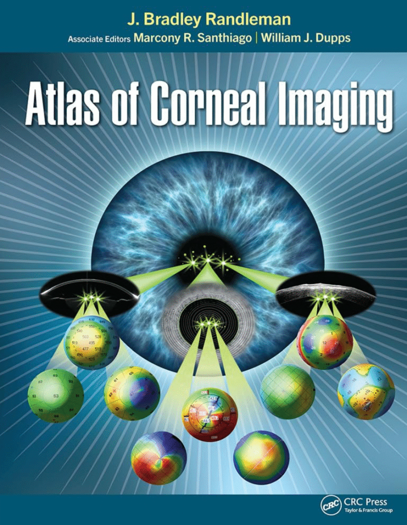
Atlas of Corneal Imaging-Understanding Corneal Imaging: A Case-Based Approach
For anyone involved in the clinical evaluation of the cornea—whether as an ophthalmologist, surgeon, or optometry trainee—having access to practical, visual resources is essential. If you’re looking to explore how corneal imaging works across different devices and technologies, and you want to build a deeper understanding of how subtle corneal pathologies present in real cases, Atlas of Corneal Imaging is a valuable text to consider.
This comprehensive atlas, authored by Drs. J. Bradley Randleman, Marcony Santhiago, and William J. Dupps Jr., offers a visual-first approach to corneal diagnostics. With more than 1200 images and illustrations drawn from real clinical cases, the book serves as a reference for those seeking to interpret imaging findings with greater clarity and precision.

Get the book via Amazon: Atlas of Corneal Imaging-Understanding Corneal Imaging
What You Can Expect to Learn
If you’re studying corneal imaging and want to understand how topography, tomography, and other technologies are applied in practice, this book guides you through:
- Basic interpretation of topographic maps
- Evaluation of corneal ectasia and keratoconus
- Preoperative and postoperative assessments for refractive and cataract surgery
- Complications in corneal and refractive surgery
- Clinical findings and how they appear across different imaging modalities
Each case is presented with multiple images from the same eye using different technologies, helping readers compare and correlate findings across platforms.
Why This Approach Stands Out
One of the challenges in clinical eye care is recognizing subtle abnormalities before they progress into significant pathology. This resource emphasizes how to detect early signs of corneal weakening—such as in keratoconus or postoperative ectasia—using a wide range of imaging tools. By seeing how various devices represent the same pathology, readers develop the ability to identify patterns regardless of the technology they use.
Rather than focusing on a single diagnostic system, the book brings together examples from many commonly used instruments, making it broadly applicable for clinicians working with different brands or models.
About the Lead Author: Dr. J. Bradley Randleman
Dr. Randleman is widely respected in the field of cornea and refractive surgery. His work has contributed significantly to the understanding of corneal biomechanics and refractive surgery complications. He serves as Editor-in-Chief of the Journal of Refractive Surgery and has authored over 165 peer-reviewed publications. His background in both clinical and academic ophthalmology adds depth and authority to the content presented in this book.
Who Might Benefit from This Resource?
This book may be especially helpful for:
- Ophthalmology residents and fellows studying corneal imaging
- Practicing surgeons seeking to refine their interpretation of imaging data
- Optometrists who co-manage surgical patients or assess corneal health
- Educators looking for visual teaching aids in cornea and anterior segment
Final Note
If you are currently learning how to analyze corneal imaging data or looking to strengthen your clinical interpretations with the help of case-based visuals, Atlas of Corneal Imaging offers a structured, image-rich resource to support your study.
You can read more about the book or find it through trusted academic booksellers and platforms like Amazon.
Read MORe articles
