
What Is Persistent Pupillary Membrane (PPM)?
A Clinical Insight With a Real Case Example of Persistent Pupillary Membrane
During a routine eye examination, certain findings may appear unusual yet represent normal variations of ocular development. Persistent Pupillary Membrane (PPM) is one such condition—frequently harmless, often misunderstood, and visually striking when seen under slit-lamp magnification.
This article explains what PPM is, why it occurs, how it appears clinically, and includes a documented paediatric case to provide real-world understanding.
Understanding Persistent Pupillary Membrane
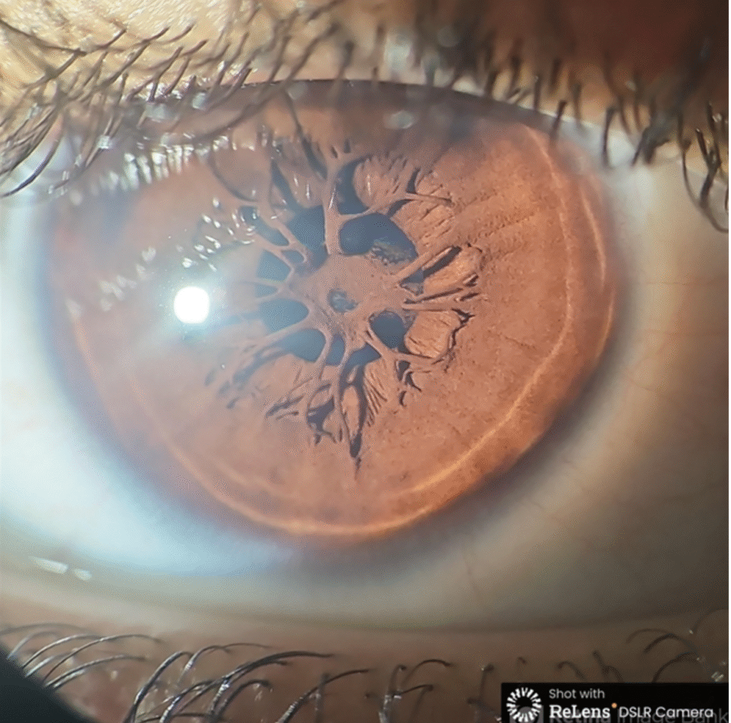
Persistent Pupillary Membrane (PPM) consists of thin, fibrous strands crossing the pupil. These strands are remnants of a fetal vascular membrane that supplied nutrients to the developing crystalline lens during intrauterine life.
Under normal circumstances, this membrane regresses completely before birth. When partial regression occurs, the remaining tissue persists into childhood or adulthood as PPM.
Is Persistent Pupillary Membrane Common?
Yes. Mild forms of PPM are relatively common and are seen in a significant percentage of the population during slit-lamp examination. Most individuals are completely unaware of its presence.
Key points:
- Congenital in origin
- Non-progressive
- Usually asymptomatic
- Often detected incidentally
Clinical Appearance of PPM
On slit-lamp biomicroscopy, PPM appears as:
- Fine, thread-like or web-like strands
- Extending from one part of the iris to another
- Sometimes attached to the pupillary margin
- Rarely dense enough to obscure the visual axis
The appearance may vary from barely visible strands to more prominent membranes, depending on the extent of persistence.
Clinical Case Example: Persistent Pupillary Membrane in a Child
Case Description
A 10-year-old female patient underwent slit-lamp examination, during which persistent pupillary membrane strandswere observed crossing the pupil.
Examination Findings
- Fine membranous strands arising from the iris
- No significant obstruction of the visual axis
- No associated inflammation or pathology
- Visual function preserved
This presentation represents a classic benign form of PPM, requiring observation rather than intervention.
Role of Slit-Lamp Photography in This Case
High-resolution slit-lamp imaging was instrumental in documenting the condition clearly. The photograph was captured using a slit lamp biomicroscope with a smartphone camera, demonstrating how accessible technology can still yield clinically valuable documentation.
Such images are essential for:
- Teaching and academic reference
- Patient and parent counselling
- Differentiation from pathological membranes
When Can PPM Affect Vision?
Although rare, vision may be affected when:
- The membrane is thick or centrally located
- Multiple strands form a dense network
- It is associated with other ocular anomalies
In these cases, treatment options such as Nd:YAG laser membranotomy or surgical removal may be considered, particularly in children at risk of amblyopia.
Management and Prognosis
For most patients:
- No treatment is required
- Routine follow-up is sufficient
- Prognosis is excellent
Parental reassurance is a key component of management in paediatric cases.
Why Understanding PPM Matters
PPM is often mistaken for pathological conditions by patients and non-specialists. Proper understanding prevents unnecessary anxiety and interventions. Clinically, it also highlights the importance of recognising normal developmental remnants versus disease.
Image Credit and Source
Photographer:
Dr. Luai Abu-Ismail
Ophthalmology Department, Islamic Hospital
Imaging Device:
Slit lamp biomicroscope photograph captured using a smartphone camera
Patient Details:
10-year-old female with persistent pupillary membrane
Image Source:
American Society of Retina Specialists (ASRS) Image Bank
https://imagebank.asrs.org/file/133950/persistent-pupillary-membrane-ppm
Conclusion
Persistent Pupillary Membrane is a benign congenital finding that reflects incomplete regression of fetal ocular structures. While its appearance may seem dramatic under magnification, it is usually harmless and requires only observation.