Skip to content
Optometryskills

What is Calcific Band Keratopathy?

November 5, 2025
Calcific Band Keratopathy
Index

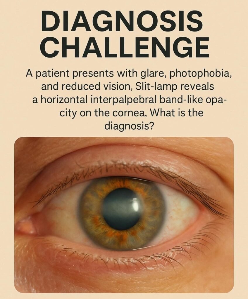
    Calcific Band Keratopathy: Key Clinical Insights for Optometrists

    What is Calcific Band Keratopathy?

    Calcific Band Keratopathy (CBK) is a degenerative corneal condition characterised by calcium hydroxyapatite deposition in the:

    • Bowman’s layer
    • Epithelial basement membrane
    • Superficial anterior stroma

    The deposits form a horizontal, interpalpebral, band-like opacity across the cornea — hence the name band keratopathy.


    Why It Matters for Optometrists

    Optometrists are often the first point of contact for these patients. Recognising CBK enables timely referral, improves comfort, and prevents visual decline.


    Clinical Presentation

    Symptoms

    • Decreased visual acuity (gradual)
    • Glare & photophobia
    • Foreign-body sensation
    • Tearing or irritation

    Signs on Slit-Lamp

    • Horizontal band opacity across interpalpebral zone
    • Begins at 3 and 9 o’clock and spreads centrally
    • Surface may appear rough and plaque-like
    • Lucid interval at limbus
    • Advanced cases: epithelial breakdown, pain

    Pearl: CBK typically spares the superior and inferior cornea, as they are protected by the eyelids.


    Differential Diagnosis

    ConditionKey Difference
    Salzmann’s Nodular DegenerationElevated bluish-white nodules
    Lipid KeratopathyAssociated vascularisation, lipid deposits
    Spheroidal DegenerationGolden, oily granules; pinguecula-like
    Band-shaped Keratopathy post traumaLocalised to area of previous injury

    Etiology & Associations

    CBK may be idiopathic but commonly associated with:

    Ocular Causes

    • Chronic uveitis (e.g., juvenile idiopathic arthritis)
    • Chronic corneal edema
    • Long-standing glaucoma
    • Keratoconjunctivitis sicca
    • Silicone oil after retinal surgery
    • Phthisical eye

    Systemic Causes

    • Hypercalcemia (e.g., hyperparathyroidism)
    • Chronic kidney disease
    • Sarcoidosis
    • Vitamin D toxicity

    Clinical Reminder: Always evaluate systemic history when CBK is detected.


    Investigations

    For new or bilateral unexplained cases:

    • Serum calcium & phosphate
    • Parathyroid hormone levels
    • Renal function tests

    Ocular assessment:

    • Slit-lamp magnification & optic section
    • Fluorescein staining: highlights irregular surface

    Management

    Non-Surgical / Supportive

    • Lubricating drops/ointments
    • Bandage contact lenses for pain
    • Avoid calcium-containing topical meds if possible

    Definitive Treatment

    TreatmentNotes
    EDTA chelation (gold standard)Removes calcium; often excellent visual recovery
    Superficial keratectomyFor deeper or dense plaques
    PTK (Phototherapeutic Keratectomy)If recurrence or deeper stromal involvement

    Referral: Refer to a corneal specialist for EDTA chelation/surgical removal.


    Prognosis

    • Vision often improves significantly post-treatment
    • Recurrence possible, especially if underlying cause persists

    Follow-Up Advice:

    • Monitor ocular inflammation
    • Support lubrication
    • Address systemic conditions

    Quick Summary for Students

    FeatureDescription
    LocationInterpalpebral zone, Bowman’s layer
    AppearanceWhite-grey, band-like plaque
    SymptomsGlare, photophobia, decreased vision
    Key AssociationsChronic uveitis, renal disease, hypercalcemia
    TreatmentEDTA chelation +/- PTK

    Optometrist’s Take-Home Message

    CBK is easy to overlook in early stages. Every slit-lamp exam is an opportunity to protect vision. Remember:

    • Look for band-like calcium at 3 & 9 o’clock
    • Ask about systemic & inflammatory conditions
    • Provide early referral for chelation therapy
    • Support the ocular surface to improve comfort

    With proper recognition and timely intervention, patients can enjoy significant improvement in clarity and comfort.

    Calcific Band Keratopathy