
Understanding Key Signs in Rhegmatogenous Retinal Detachment
Rhegmatogenous retinal detachment (RRD) is the most common and vision-threatening type of retinal detachment. The word rhegmatogenous comes from the Greek term rhegma, meaning “break” or “fissure.” This name highlights the underlying mechanism — a full-thickness break in the neurosensory retina that allows liquefied vitreous to seep underneath, separating it from the retinal pigment epithelium (RPE).
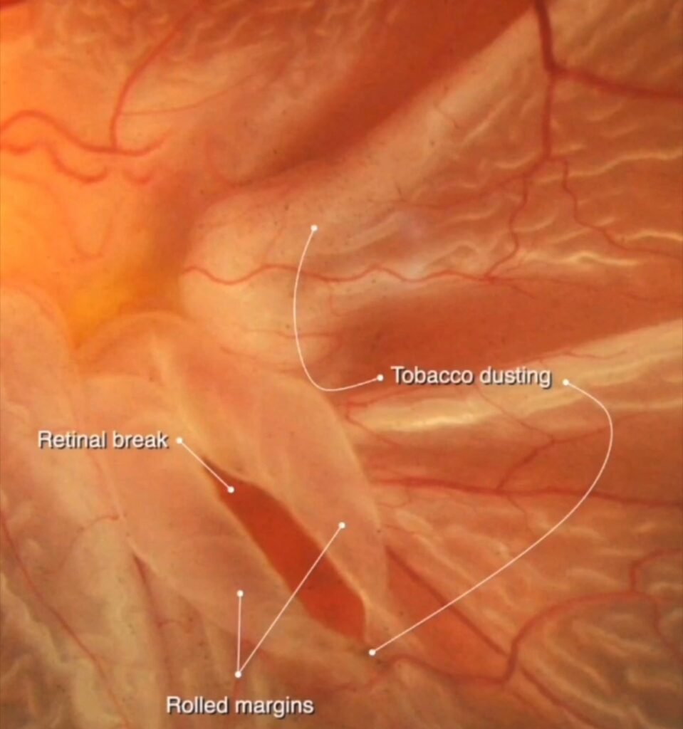
Recognizing the classic signs of a retinal break, rolled margins, and tobacco dusting is crucial for early diagnosis and prompt referral, which can often make the difference between vision restoration and permanent visual loss.
1. Retinal Break
A retinal break is the initiating event in rhegmatogenous retinal detachment. It refers to a full-thickness defect in the sensory retina, through which liquefied vitreous gains access to the subretinal space.
These breaks may take the form of tears, holes, or dialyses, depending on the cause. In most cases, posterior vitreous detachment exerts traction on the retina, especially at weak points such as lattice degeneration, leading to a tear.
On fundus examination, a retinal break appears as a reddish, irregular slit against the pale detached retina. The margins are often lifted and distinct, marking the point of fluid entry beneath the retina. Identifying this break is vital, as sealing it (usually with laser photocoagulation or cryotherapy) forms the basis of surgical management.
2. Rolled Margins
When a retinal tear forms, the edges of the retina tend to curl inward due to vitreoretinal traction. These rolled margins are characteristic of rhegmatogenous retinal detachment.
The rolling of the retinal flap occurs as liquefied vitreous fluid enters beneath the retina, pushing it away from the underlying RPE. The detached portion becomes mobile and undulating, and the rolled edges appear thickened and elevated.
The presence of rolled margins helps distinguish rhegmatogenous retinal detachment from tractional or exudative types, where such a tear and curling of margins are absent. Clinically, this finding indicates that the retina has lifted off and is freely mobile, emphasizing the urgency of surgical repair.
3. Tobacco Dusting (Shafer’s Sign)
One of the earliest and most pathognomonic signs of a retinal break is tobacco dusting, also known as Shafer’s sign. It appears as fine brown pigment granules suspended in the vitreous cavity, resembling tobacco dust particles floating in light.
These pigments originate from retinal pigment epithelial cells that get dislodged when a retinal tear occurs. The presence of this dust in the anterior vitreous is a strong indicator of a recent retinal break or detachment.
Shafer’s sign can be observed during slit-lamp examination with a +90D lens or during indirect ophthalmoscopy. Its detection is particularly important in patients who present with symptoms of flashes, floaters, or sudden vision loss, as it alerts the clinician to search carefully for a small or peripheral retinal break.
Clinical Significance
Recognizing these three findings — retinal break, rolled margins, and tobacco dusting — forms the cornerstone of diagnosing rhegmatogenous retinal detachment.