
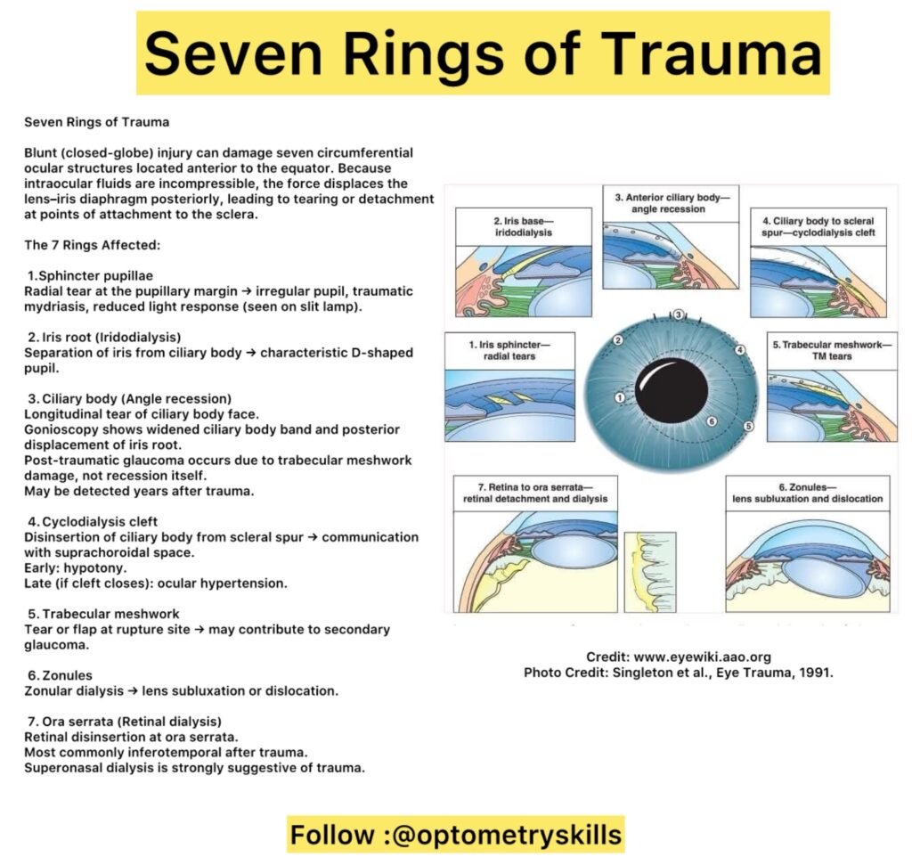
Seven Rings of Trauma in Blunt Ocular Injury
Blunt trauma to the eye can cause a characteristic pattern of damage known as the “Seven Rings of Trauma.” These rings refer to seven circumferentially oriented ocular structures located anterior to the equator of the globe that are particularly vulnerable during a closed-globe injury.
When the eye is struck by a blunt force, the intraocular fluids—being incompressible—transmit the energy throughout the globe. This sudden force pushes the lens–iris diaphragm posteriorly. Because these tissues are firmly attached to the scleral wall at specific points, the transmitted energy can cause splitting, tearing, or detachment at these attachment sites. The result is a predictable series of injuries involving seven anatomical structures.
Understanding these rings is essential for clinicians, as some findings may appear immediately after trauma, while others—such as secondary glaucoma—may develop years later.
1. Sphincter Pupillae
Blunt trauma may cause a radial tear at the pupillary margin, visible on slit-lamp examination. Clinically, this may present as:
An irregular or distorted pupil Reduced or absent light reflex Traumatic mydriasis
2. Iris Root – Iridodialysis
Iridodialysis refers to the separation of the iris root from the ciliary body.
It often produces a characteristic “D-shaped” pupil and may be associated with glare, diplopia, or cosmetic concerns.
3. Ciliary Body – Angle Recession
Angle recession is a longitudinal tear of the ciliary body face, separating circular from longitudinal muscle fibers. On gonioscopy, it appears as:
Widened ciliary body band Posterior displacement of the iris root
Importantly, post-traumatic glaucoma is not caused by the recession itself but by associated damage to the trabecular meshwork. Angle recession is one of the most common findings after blunt contusion and may still be visible years after the initial injury.
4. Cyclodialysis Cleft
A cyclodialysis cleft occurs when the ciliary body detaches from the scleral spur, creating an abnormal communication between the anterior chamber and the suprachoroidal space.
Early complication: Hypotony due to excessive aqueous outflow Late complication: Ocular hypertension if the cleft closes with impaired aqueous drainage
5. Trabecular Meshwork
Blunt trauma may cause a tear or flap in the trabecular meshwork, contributing to impaired aqueous outflow and secondary glaucoma.
6. Zonules
Damage to the zonular fibers may result in zonular dialysis, leading to:
Lens subluxation Complete lens dislocation
This can significantly affect visual acuity and may require surgical intervention.
7. Ora Serrata – Retinal Dialysis
Retinal dialysis is the disinsertion of the retina at the ora serrata.
Most commonly affects the inferotemporal quadrant after trauma Superonasal dialysis is strongly suggestive of traumatic origin
This condition can lead to retinal detachment if not diagnosed and managed promptly.
Clinical Significance
The Seven Rings of Trauma provide a systematic framework for evaluating patients with blunt ocular injury. A comprehensive examination—including slit-lamp biomicroscopy and gonioscopy—is essential. Long-term follow-up is equally important, as complications such as secondary glaucoma may appear years after the initial trauma.
Recognizing these injury patterns allows for early intervention and better visual prognosis.
Credit: EyeWiki (www.eyewiki, aao.org)
Photo Credit: Singleton et al., Eye Trauma, 1991.