
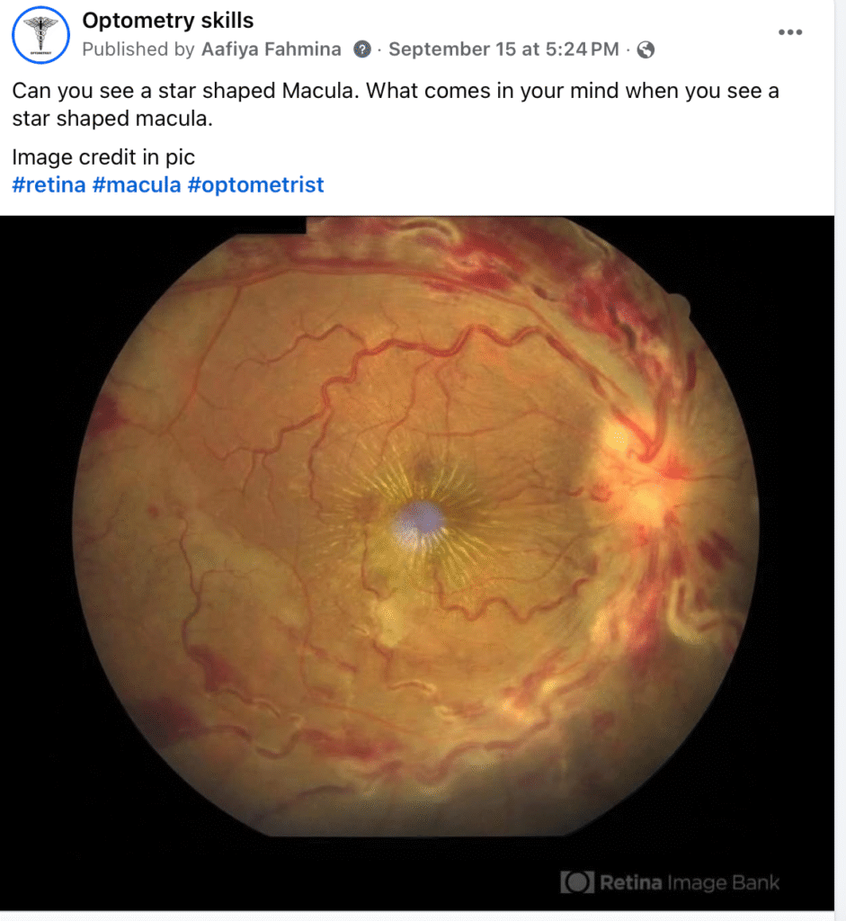
Differential Diagnosis of Macular star
Answer: Central Retinal Vein Occlusion (CRVO)
A star-shaped macula immediately raises suspicion for lipid exudates deposited in Henle’s layer, which is seen in:
• Neuroretinitis especially from Cat Scratch Disease
• Hypertensive retinopathy
• Papillitis / Papilledema with exudation
• Retinal vascular occlusions (including CRVO)
In this case, the dilated tortuous veins, Diffuse intraretinal hemorrhages in all quadrants (“blood and thunder” appearance), and macular star point strongly toward Central Retinal Vein Occlusion (CRVO) with macular edema and hard exudates.
![]() So the “star” isn’t exclusive to one disease — but in the presence of venous engorgement and hemorrhages, CRVO becomes the top differential.
So the “star” isn’t exclusive to one disease — but in the presence of venous engorgement and hemorrhages, CRVO becomes the top differential.
Differential Diagnosis of a Star-Shaped Macula
• Neuroretinitis (Cat scratch, TB, syphilis, viral, idiopathic)
Signs: Disc edema + macular star, Few or no retinal hemorrhages,Often young patients with history of fever, lymphadenopathy, or exposure to cats
• Hypertensive retinopathy (malignant HTN)
Signs: Arteriolar narrowing, cotton wool spots, flame hemorrhages, disc edema, macular star, Patient usually has severe uncontrolled BP
• CRVO
Signs: Dilated, tortuous retinal veins + extensive flame/blot hemorrhages + macular edema + hard exudates (sometimes star-shaped), Patient usually is of Older age or has vascular risk factors (HTN, DM, hyperlipidemia)
++Note : CRVO is usually a unilateral, sectoral problem related to a vein blockage, while hypertensive retinopathy is typically bilateral and involves the arterial system