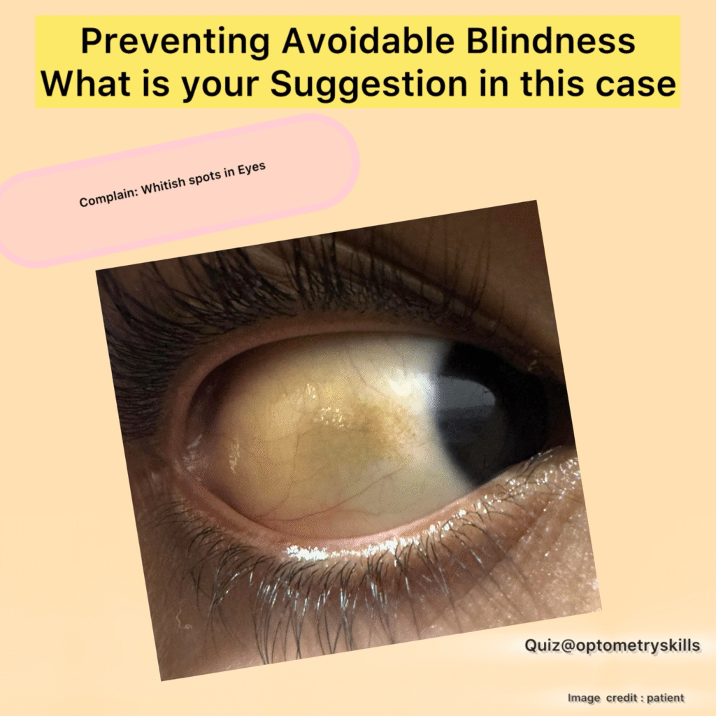
Conjunctival Naevus
Conjunctival naevi are the most common benign pigmented lesions found on the ocular surface, frequently appearing during childhood or adolescence. While typically harmless, optometrists play a crucial role in distinguishing these benign lesions from more serious conditions like conjunctival melanoma. Careful observation, documentation, and timely referral are key to optimal patient care.
What is a Conjunctival Naevus?

A conjunctival naevus is a pigmented, melanocytic lesion located on the bulbar conjunctiva, often near the limbus, caruncle, or plica semilunaris. Most remain stable throughout life, but changes in appearance or symptoms should always prompt a thorough evaluation.
Optometric Management Guidelines
See also Diagnosing Hypertensive Retinopathy
Slit-Lamp Examination: Document size, color, elevation, and vascular patterns. Photography: Capture high-resolution baseline photos for comparison. Differential Diagnosis: Rule out melanoma, primary acquired melanosis (PAM), or racial melanosis.
2. Identifying Benign vs Suspicious Features
- Benign Features
- Suspicious Features
- Stable appearance over time
- Rapid growth or size increase
- Presence of intralesional cysts
- Loss of cysts or sudden change in structure
- Uniform pigmentation and smooth margins
- Irregular pigmentation or feeder vessels
- No associated symptoms
- Discomfort, redness, irritation, or bleeding
| Benign Features | Suspicious Features |
|---|---|
| Stable appearance over time | Rapid growth or size increase |
| Presence of intralesional cysts | Loss of cysts or sudden change in structure |
| Uniform pigmentation and smooth margins | Irregular pigmentation or feeder vessels |
| No associated symptoms | Discomfort, redness, irritation, or bleeding |
3. Follow-Up Recommendations
Benign, stable naevi: Monitor annually with repeat photos. Subtle changes (size or color): Follow up in 3-6 months. Concerning signs: Prompt referral to ophthalmology or ocular oncology for further evaluation.
See also What is Mixed Astigmatism?
When to Refer
Optometrists should refer urgently if:
There is rapid growth New feeder vessels appear The lesion loses cystic characteristics The patient reports symptoms like irritation or redness
Ophthalmologists may consider surgical excision or biopsy if malignancy cannot be ruled out.
Patient Education
Educating patients is crucial:
Reassure about the benign nature of most naevi. Explain the importance of yearly monitoring. Encourage patients to report any changes in size, shape, or color.
Conclusion
Conjunctival naevi are common and usually harmless, but careful observation ensures early detection of atypical changes. As frontline eye care providers, optometrists must confidently monitor, document, and refer when necessary, safeguarding ocular health and providing peace of mind to patients.
