
Can a Worm Really Live in the Human Eye? Understanding Ocular Filariasis
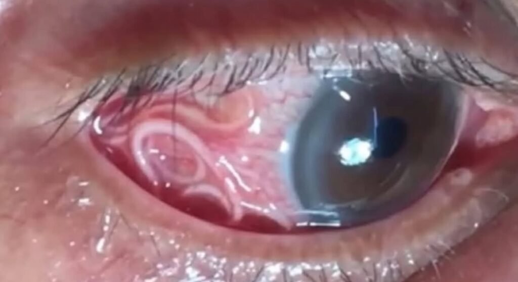
Ocular filariasis is a rare but alarming parasitic infection that affects the human eye. Caused by filarial nematodes (microscopic worms), this condition can lead to visible worms moving beneath the eye’s surface, intense irritation, and swelling.
While it is uncommon, especially outside endemic regions, ocular filariasis can cause serious discomfort and potential vision problems if not treated promptly.
Causes of Ocular Filariasis
Ocular filariasis is caused by the transmission of parasitic worms through insect bites. The main species responsible include:
Loa loa (African Eye Worm): Transmitted through the bite of the Chrysops (deer or mango fly), this parasite migrates through the subcutaneous tissues and can eventually enter the eye.
Dirofilaria species: Typically found in animals, these worms occasionally infect humans (a zoonotic infection). They can lodge in the eyelid or under the conjunctiva, forming nodules or becoming visible through the sclera.
Other Filarial Worms: Infections from Brugia malayi, Onchocerca volvulus, or Wuchereria bancrofti may also involve the eyes, especially in severe systemic infections.
Symptoms of Ocular Filariasis
Patients may present with a variety of eye symptoms depending on the worm’s location and movement within the eye:
Visible Worm: A live, motile worm may be seen wriggling under the conjunctiva or moving across the eye’s surface.
Lid Edema and Chemosis: Swelling of the eyelid and the conjunctiva due to inflammation or allergic reaction to the parasite.
Inflammation: In some cases, anterior uveitis and corneal edema can occur, causing redness, pain, and blurred vision.
Subconjunctival or Eyelid Nodules: The parasite may form encapsulated nodules as it grows or becomes trapped. Other Symptoms: Persistent itching, irritation, tearing, and a sensation of a foreign body in the eye are common complaints.
Diagnosis
Diagnosis relies on both clinical and laboratory investigations:
Clinical Examination: Direct visualization of the worm beneath the conjunctiva or inside the anterior chamber is often diagnostic. Blood Smears: In cases of Loa loa infection, microfilariae (larval forms) can be detected in peripheral blood, particularly during the daytime when they are most active.
Treatment and Management
Surgical Removal: The primary and most effective treatment for ocular filariasis is the surgical extraction of the worm under local anesthesia. This provides immediate relief and prevents further eye damage.
Medication: Diethylcarbamazine (DEC): Commonly used to treat Loa loa and other filarial infections, DEC helps eliminate microfilariae and adult worms from the bloodstream.
Ivermectin or Albendazole: In some cases, these antiparasitic drugs may be prescribed.
Anti-inflammatory Therapy: Topical corticosteroids such as prednisolone eye drops help control inflammation and prevent post-surgical irritation.
Note : This is not a clinical advice but a case discussion for Optometrist for study purpose.
Conclusion
Though rare, ocular filariasis serves as a reminder of the intricate relationship between parasitic infections and eye health. Prompt medical attention, surgical intervention, and appropriate medication can lead to full recovery without long-term damage.
Awareness among healthcare professionals and individuals living in endemic regions is vital for early recognition and prevention of this unusual yet significant condition.
More reading resource: Ocular Filariasis:
Centers for Disease Control and Prevention (CDC) – Loiasis (African Eye Worm) CDC Dirofilariasis Information – Dirofilariasis – Parasites – DPDx American Academy of Ophthalmology (AAO) – Ocular Parasitosis StatPearls (NCBI Bookshelf) – Filariasis