
Optociliary Shunt Vessels: Overview, Etiology, Diagnosis, and Management
Overview
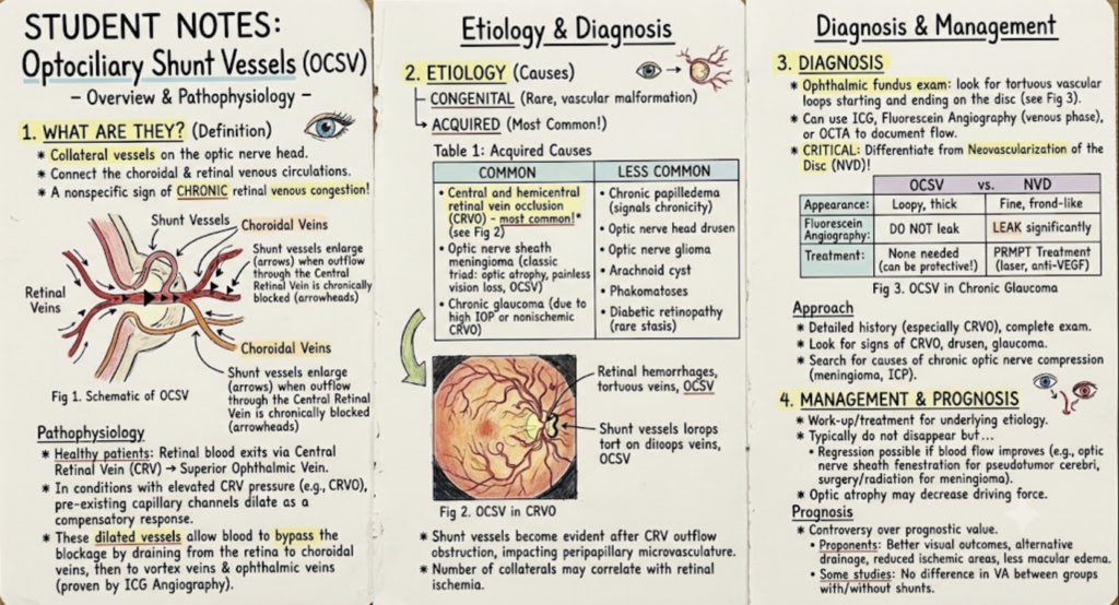
Optociliary shunt vessels, also referred to as retinochoroidal shunt vessels of the optic disc or retinochoroidal venous collaterals, are collateral vessels located on the optic nerve head that directly connect the choroidal and retinal circulations.
These vessels are associated with a range of ophthalmic and systemic conditions, including central retinal vein occlusion, optic nerve sheath meningioma, chronic glaucoma, and chronic papilledema. Pathologically, they represent a non-specific clinical sign of chronic retinal venous congestion.
Pathophysiology
In a standard physiological setting, venous blood from the retina leaves the eye through the central retinal vein (CRV), which traverses the center of the optic nerve to drain into the superior ophthalmic vein.
When the normal outflow through the central retinal vein is chronically obstructed, pre-existing anastomotic capillary channels gradually dilate and enlarge in response to elevated intravascular pressure. These resulting optociliary shunt vessels serve as alternative venous pathways that redirect blood from the retinal venous circulation to the peripapillary choroidal veins in the pre-laminar region of the optic nerve.
Studies using Indocyanine Green Angiography (ICG) have demonstrated that these dilated collateral channels allow retinal venous blood to bypass the blocked central retinal vein, exiting the orbit via:
- The choroidal circulation
- Vortex veins
- Ophthalmic veins
Note: Regression of these shunt vessels has been documented following procedures such as optic nerve sheath fenestration, which relieves pressure on the venous system.
Etiology
Optociliary shunt vessels are categorized based on whether they are congenital or acquired.
1. Congenital
- Rarity: Congenital cases are rare.
- Mechanism: They represent a developmental vascular malformation connecting the retinal and choroidal venous circulations.
2. Acquired
Acquired vessels develop secondary to ophthalmic conditions that cause impaired retinal venous outflow. The dilation of retinochoroidal vessels usually becomes evident following central retinal vein outflow obstruction, impacting the peripapillary microvascular network. Additionally, the sheer number of collaterals present may correlate with the severity of retinal ischemia.
| Cause Frequency | Ophthalmic / Systemic Conditions |
| Common Causes | • Central and Hemicentral Retinal Vein Occlusion (CRVO): Most common cause overall. • Optic Nerve Sheath Meningioma: Forms a classic triad (Optic atrophy + Painless visual loss + Optociliary shunt vessels). • Chronic Glaucoma: Caused either by high IOP/optic cup distortion distorting retinal veins, or a prior, unrecorded non-ischemic CRVO. |
| Less Common Causes | • Chronic papilledema / Post-papilledema optic atrophy • Optic nerve head drusen • Optic nerve glioma • Arachnoid cyst of the optic nerve • Phakomatoses • Diabetic retinopathy (secondary to microangiopathy and venous stasis) |
Diagnosis
On a standard ophthalmic fundus examination, optociliary shunt vessels typically appear as tortuous, loopy vessels that start and end on the optic disc.
Diagnostic Modalities
Flow within these shunt vessels can be visualized and documented using:
- Indocyanine Green Angiography (ICG)
- Fluorescein Angiography (specifically during the venous phase)
- Optical Coherence Tomography Angiography (OCTA)
Differential Diagnosis: Shunt Vessels vs. Neovascularization
A critical diagnostic step is differentiating optociliary shunt vessels from Neovascularization of the Disc (NVD). Misdiagnosis can lead to inappropriate management.
| Feature | Optociliary Shunt Vessels (OCSV) | Neovascularization of the Disc (NVD) |
| Appearance | Loopy, thick, tortuous | Fine, fragile, frond-like network |
| Fluorescein Angiography | No leakage | Profuse leakage |
| Pathological Role | Compensatory / Protective against ischemia | Pathologic / High risk of vitreous hemorrhage |
| Treatment Required | None directed at the vessels | Prompt laser photocoagulation or anti-VEGF injections |
Clinical Approach
- History: Take a detailed history with focus on prior central retinal vein occlusions.
- Fundus Examination: Look for underlying structural causes such as disc cupping (glaucoma), drusen, or venous occlusion changes.
- Neuro-Ophthalmic Workup: If retinal causes are absent, evaluate for chronic optic nerve compression (e.g., optic nerve sheath meningioma) or idiopathic intracranial hypertension. In papilledema, the presence of these vessels helps confirm that the elevated intracranial pressure is chronic.
Management and Prognosis
Management
Treatment strategies do not target the shunt vessels directly, as they do not leak or pose an inherent danger. Instead, management is focused entirely on treating the underlying etiology.
Although these collaterals traditionally persist, they may decrease in caliber or undergo complete regression if blood flow through the central retinal vein improves. Regression has been documented in:
- Pseudotumor Cerebri: Following successful optic nerve sheath fenestration.
- Optic Nerve Sheath Meningioma: Spontaneously, post-surgical resection, or following radiation therapy (which may resolve vessels immediately or over several years).
- Optic Atrophy: As nerve atrophy progresses, the diminished structural mass may reduce venous obstruction and driving pressure, leading to vessel involution.
Prognostic Value
The overall prognostic value of retinochoroidal collateral vessels remains a point of clinical debate:
- Proposed Benefits: Some evidence suggests collaterals lead to better visual outcomes and lower rates of macular edema by providing an alternative venous drainage path that reduces ischemic areas.
- Alternative Findings: Other studies report no significant difference in long-term visual acuity outcomes between patients with and without optociliary shunt vessels.

Optometry education for Optometry students and eye care professionals. Managed by qualified optometrists. For informational purposes only — not a substitute for professional eye care.